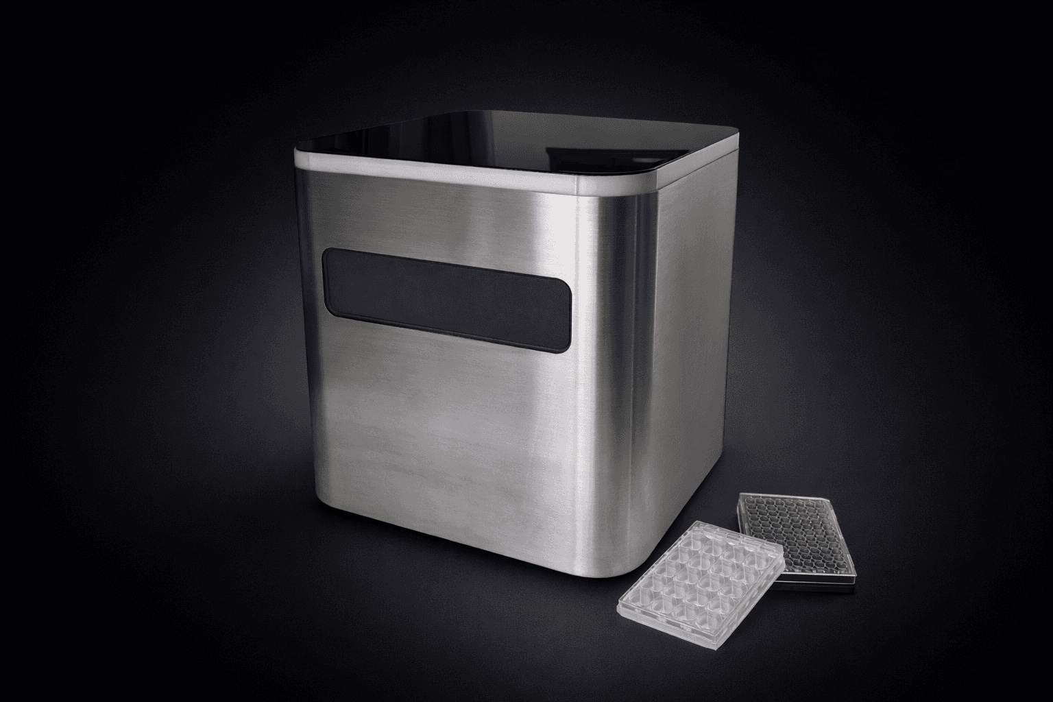
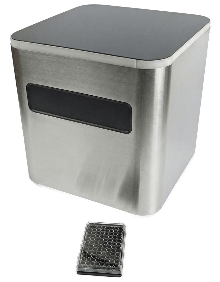
CCA Microscope
Automated live-cell imaging with integrated environmental control for long-horizon culture monitoring. Capture confluency and morphology trends over days or weeks without moving plates between instruments. When connected to CCA Labs, each image and metric is linked to protocol step, sample, and timepoint.
Built for Time-Resolved Cell Biology
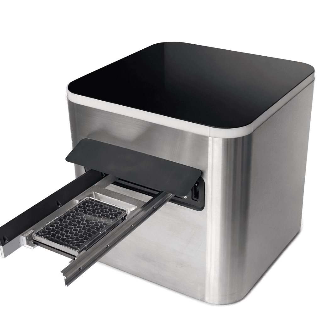
Integrated incubation, precision optics, and automated plate scanning in a compact benchtop footprint. Designed for biosafety cabinet workflows and robotic plate handoff where deterministic timing matters.
Incubation and Imaging in One Time-Aligned System
Incubation and imaging in one unit for longitudinal live-cell monitoring. Cells stay under controlled conditions while the system scans a single SBS plate on a schedule. When connected to CCA Labs, images, acquisition settings, and results are linked to the protocol, sample, and timepoint for traceability.
- Built-in incubator with dynamic control of temperature, CO₂, and O₂
- Hypoxia support (1–21% O₂) for tumor and microenvironment workflows
- Benchtop footprint designed for biosafety cabinets and robot access
- Remote monitoring and management via CCA Labs (optional cloud connection)
- Quantitative outputs (confluency, morphology) with deviation alerts
- Native REST API for integration and protocol-triggered automation workflows
What's Inside
Optical System
Automation
Incubator
Software & Analysis
Connectivity
Physical
How We Compare
Side-by-side comparison against common live-cell imaging setups used in research labs.
Feature
CCA Microscope
Typical Competitor
Benchtop Design
Fits in laminar flow hoods, integrates with liquid handling robots
Integrated Environmental Control
Built-in temp, CO₂, O₂, humidity control with time-varying profiles
Robotic Integration
Motorized tray for easy liquid handling and robotic arm integration
Native Hypoxia Support
Hypoxic conditions control 0-21%
Embedded Metadata
Protocol-linked, environment-synced, no CSV exports
Native REST API
Programmatic access for reproducible analysis workflows
Built-In Quantitative Analysis
Confluency, morphology, and viability metrics at acquisition time
Software Licensing
All software included, no per-seat fees
Benchtop Integration
Compact footprint fits inside laminar flow hoods and supports robotic plate handoff. This enables repeatable timing for feeding, seeding, and passaging in constrained lab space.
Integrated Mini-Incubator
Built-in control of temperature, CO₂, O₂, and humidity reduces transfer steps between instruments. Time-varying gas and temperature profiles support hypoxia and stress-response workflows.
Protocol-Linked Records
Each image is linked to protocol step, sample identity, and environmental context. Time and metadata stay attached to the record, which simplifies run review and comparison.
Native API and Analysis Pipelines
REST API access supports scripted analysis and integration with existing lab software. Image storage, processing, and model outputs can be managed without manual file stitching.
Built-In Quantitative Analysis
Built-in models report confluency trajectories by timepoint, morphology flags, and viability indicators on scheduled acquisitions. These outputs can be used as review checkpoints or as trigger signals in rule-defined workflows.
Procurement Clarity
Hardware, software, installation, and training are scoped together in procurement discussions. Academic collaboration programs are handled through direct contact.
Design Details

Closed Configuration
Automated Plate Loading
Built for Longitudinal Cell Work
CCA Microscope combines incubation and imaging in one unit to capture consistent time-series data on a single SBS plate. Quantitative readouts support confluency, morphology, and growth trends over time. Today, it guides manual interventions; dALI extends the same workflow definitions to threshold-triggered execution.
iPSC & Stem Cell Workflows
iPSC, MSC, primary adherent cellsMaintenance & Expansion
Track colony morphology and growth over days. Use defined imaging checkpoints for colony selection and to time feeds and passages consistently.
Differentiation Monitoring
Capture stage-dependent morphology changes with time-series imaging. Define checkpoints and compare runs with the same imaging schedule.
Quality Control
Continuous imaging provides objective evidence for drift, stress, or contamination signals without relying on intermittent manual checks.
Organoids & 3D Models
3D growth, morphogenesis, growth trackingGrowth Over Time
Track organoid growth and morphology across days with consistent timepoints, enabling comparable time-series datasets.
Developmental Dynamics
Capture changes in structure and patterning as they emerge. Time-series images provide evidence of when transitions occur.
3D Disease Models
Compare phenotypes across genotypes or conditions using matched imaging schedules and controlled incubation parameters.
Advanced Research with Dynamic Control
Normoxia ↔ hypoxia, stress profilesHypoxia Studies
Control O₂ to study hypoxia response and microenvironment effects. Compare runs under defined O₂ conditions with imaging evidence.
Condition Sweeps
Run controlled changes in incubation parameters and quantify how morphology and growth respond across timepoints.
Reproducible Time-Series
Standardised scheduling + controlled conditions reduce variability, making trends comparable across experiments and teams.
Workflows That Scale with dALI
From guided to automated executionToday: Guided Manual Steps
Use metrics to time feeding and passaging consistently. Imaging provides evidence and traceability for every intervention.
Next: Automated Execution
dALI performs liquid handling and plate movement to execute the same workflows automatically on schedule or by threshold.
Closed-Loop Decisions
Use measured state (imaging + incubation conditions) to trigger actions, creating repeatable and scalable protocol execution.
Transparent, All-Inclusive
Same instrument for academic and industrial users. Academic programs are available through direct contact, while industrial customers purchase commercially.
All prices include hardware, software, installation, and training. Annual maintenance contracts available upon request.